
Before to understand how our knee joint work, first we have to understand:-
1) where knee joint is present?
2) what are the components which made knee joint?
Let's start with some Introduction:-
- Knee joint is present in lower limb.
- Knee joint is the complex joint.
- Component involve in knee joint
- Femur (Thigh bone)
- Tibia (Leg bone)
- Patella (Knee cap)
- It's made up of 2 joint
- Tibiofemoral joint (tibia articulate with femur)
- Patellofemoral joint (patella articulate with femur)
- There are many structures which protect the knee joint like Ligaments, Capsule, Menisci
A. COMPONENTS
- PARTS:
- Proximal part: head, neck, greater trochanter, lesser trochanter, intertrochantric line, gluteal tuberosity.
- Middle part (Shaft)
- Lower part: medial condyle, lateral condyle, intercondylar fossa, central groove(patellar groove)

b) TIBIA (leg bone):Also known as shin bone
- PARTS:


- Proximal part: Medial condyle, Lateral condyle, Medial and lateral tibial plateau, Intercondylar eminence, tibial tuberosity.
- Middle part (Shaft)
- Distal part:medial mallelous
c) PATELLA (knee cap): It's an inverted triangle with it's apex directed inferiorly.
Posterior surface have :-
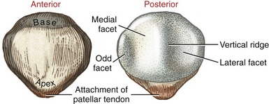
- Medial facet
- Lateral facet
- Vertical ridge
- Odd facet
B. KNEE JOINT
It involve 2 joints:-
(1) Tibiofemoral joint:
When both tibial plateau articulate with both femoral condyle it made Tibiofemoral joint.
(2) Patellofemoral joint:When posterior surface of patella articulate with central groove of femur it made Patellofemoral joint.
C. STRUCTURES
a) LIGAMENTS
1) Anterior cruciate ligament( ACL):Prevent anterior translation of tibia on femur
2) Posterior cruciate ligament(PCL): Prevent posterior ranslation of tibia on femur
3) Medial collateral ligament(MCL): Prevent valgus force (abduction)at knee
4) Lateral cruciate ligament(LCL): Prevent varus force(adduction) at knee.
 b) MENISCI
1)Medial meniscus
2)Lateral meniscus
- Act as shock absorber
- Reduce friction between tibia and femur
- Distribute weight bearing forces
c) CAPSULE
- It enclose the both tibiofemoral and patellofemoral joint.
- It's grossly composed of :-
2) thin deep synovial layer
D. MOVEMENTS
The main movement which occur at knee joint is :-

1) FLEXION:- Femur roll posteriorly and glide anteriorly on relatively fixed tibia (According to concave - convex rule)
2)EXTENSION :- Femur roll anteriorly and glide posteriorly on relatively fixed tibia (According to concave - convex rule).
 (A) Open kinematic chain( Non weight bearing)
While knee extension (last 30° of knee extension) there is lateral rotation of tibia occur on femur to complete knee extension. It's referred as Automatic rotation or Screw home mechanism.
(B) Closed kinematic chain (Weight bearing)
YOUTUBE:
D. MOVEMENTS
The main movement which occur at knee joint is :-
1. Flexion
2. Extension
2. Extension
But both Abduction/Adduction, Medial/Lateral rotation also occur in knee joint but to a lesser extent which we are unable to observe at knee joint but they are necessary for normal functioning of tibio-femoral joint.
**As we almost discuss the main components which will help us to understand the main content of this topic...." HOW OUR KNEE JOINT MOVE"(Arthrokinematic of knee joint)
TIBIOFEMORAL JOINT
a) Now here we discuss about how our knee joint work?
b) How one joint surface move on another joint surface?
c) What are the movements occur between the joint surfaces?
d) Joint surface movements depend upon:-
2) Is Convex surface is moving on concave surface or Concave surface is moving on convex surface?
(A) CLOSED KINEMATIC CHAIN(weight bearing)
When femur is moving on relatively fixed tibia. eg:- Squatting.
When femur is moving on relatively fixed tibia. eg:- Squatting.

1) FLEXION:- Femur roll posteriorly and glide anteriorly on relatively fixed tibia (According to concave - convex rule)
2)EXTENSION :- Femur roll anteriorly and glide posteriorly on relatively fixed tibia (According to concave - convex rule).
(B) OPEN KINEMATIC CHAIN( Non weight bearing)
When tibia is moving on relatively fixed femur eg :- High sitting knee flexion/Extension.
1) FLEXION :- Tibia glide and roll posteriorly on relatively fixed femur (Acc. to concave - convex rule).
2) EXTENSION :- Tibia glide and roll anteriorly on relatively fixed femur (Acc. to concave - convex rule)
SCREW - HOME MECHANISM
To complete knee flexion/Extension there is little rotation of tibia on femur occur.

While knee extension (last 30° of knee extension) there is lateral rotation of tibia occur on femur to complete knee extension. It's referred as Automatic rotation or Screw home mechanism.
Now, to flex the tibia on femur, the tibia rotate 1st medially to unlock the knee as laterally rotated tibia can't flex.
(B) Closed kinematic chain (Weight bearing)
While knee extension (last 30° of knee extension) the femur rotate medially on fixed tibia to lock the knee.
While knee flexion, initially the femur rotate laterally on fixed tibia to unlock the knee.
PATELLOFEMORAL JOINT
a) It's one of the most in-congruent joint in the body.
b) Due to in-congruence of the patella, the contact between the patella and femur changes throughout the knee Range
of motion (ROM)
c) In fully extended knee, patella lies on the patellar groove on femur.
c) In fully extended knee, patella lies on the patellar groove on femur.
d) Contact between both surface during knee movements
-Fully Extension= Only the inferior pole of the patella is making contact with femur
-10°-20° of knee flexion= Inferior margin of both the medial and lateral facets of patella
-As flexion progress(45°)=most of the part of lateral and medial facet of patella make contact
-At 90°= All portion of patella make contact with femur except odd facet
-Beyond 90° =Patella migrate inferiorly, odd facet make contact with the medial femoral condyle for the first time
-At full flexion=Patella lodge in the intercondylar groove, only lateral and odd facet make contact.


Summery:
Through this knowledge now we are able to understand how our knee joint move.
In Closed kinematic chain:-
a) Femur move on relatively fixed tibia.
b) Rolling and gliding occur in opposite direction.
c) Locking of knee, done by medial rotation of femur on tibia and unlocking done by lateral rotation of femur on tibia.
b) Rolling and gliding occur in opposite direction.
c) Locking of knee, done by medial rotation of femur on tibia and unlocking done by lateral rotation of femur on tibia.
In Open kinematic chain :-
a) Tibia move on relatively fixed femur.
b) Rolling and gliding occur in same direction.
c) Locking of knee done by lateral rotation of tibia on femur and unlocking done by medial rotation of tibia on femur.
a) Tibia move on relatively fixed femur.
b) Rolling and gliding occur in same direction.
c) Locking of knee done by lateral rotation of tibia on femur and unlocking done by medial rotation of tibia on femur.
Resources:
1) Joint structure and function, a comprehensive analysis 5th edition by Pamela K. Levangie, Cynthia C. Norkin
2) Kinesiology Of Musculoskeletal System, foundations for rehabilitation 3rd edition by Donald A. Neuman
If you want to know more you can check these link below also:
BY MANISHA GOLA (PHYSIOTHERAPIST)

5 Comments
Nice.. liked it... very knowledgeable.... this is the best way to revise the anatomy and make ur theory part more strong.. thanku for sharing... share more
ReplyDeleteGood job
ReplyDeletebest way to revise 👏
ReplyDeleteSimplified yet comprehensive. Every aspect is covered. In this current era of students try out to learn bat and cat without actually knowing the alphabets, it’s a valid attempt signifying the importance of anatomy and the biomechanics before trying out the fantasy of manipulation chiropractic osteopath techniques. I feel complete that my teachings had some effects on my students. Feeling proud for your valid attempt. Keep going. Miles to go before sleep.
ReplyDeleteKnee joint simplified ��
ReplyDeletePlease do not enter any spam link in the comment box