Introduction
Radiology is vital nearly for every sector of health care, including Physiotherapy, as it helps to confirm the provisional diagnosis which a therapist makes after a thorough clinical examination of a patient.
In this blog, we are going to study that there are so many investigations comes under radiology, but for physiotherapist, we are mostly interested in X-ray, CT and MRI, that’s why we have started this blog series and in this blog, we are going to discuss:
- Explanation of the basic properties of X rays, how it’s used to produce an image of the body organ.
- A thorough discussion about: why bone appears white and air looks black in colour.
- In the last, we would clear some important characteristics of these densities, which help us to interpret the x-ray image.
What is Radiology?
- Radiology is a branch of medicine that deals with radiant energy to diagnosis and treatment of the disease.
- It includes a broad spectrum of imaging techniques, the basic principle of imaging, however, remains the anatomical demonstration of a particular region and related abnormalities, the principle imaging modalities in which physiotherapist show interest are X-ray, CT and MRI.
Firstly, we are going to understand what is x-rays, how it produces images and fundamental concepts which are used to interpret the x-ray images.
What is the X Rays?
- X-ray is ionizing radiation: which means X rays can produce ions (charged particles) in the matter and if this ionization occurs in a tissue, it can lead to cellular damage.
- The X-ray can penetrate: but not through all the material, it can penetrate through less density matter and high-density matter can absorb it.
How X-ray images are produced?
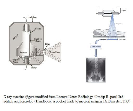
X rays are produced in the x-ray machine and are exposed to the patient body or structure that we need to see and strike the photosensitive surface(x-ray detector), based on how much x rays reaches the detector after passing through patient, a shadowgram is formed of varying intensities of a grey colour.
Radiographic Densities:
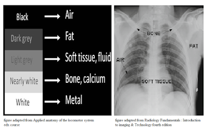
There are five basic densities in the x-ray images based on the densities of the tissues present in our body.Tissues densities are based on the following characteristics:
1) Atomic Number: higher the atomic number of a matter, the more x rays will be blocked from reaching the film. In the body minerals like calcium has the high atomic number that’s why they absorb x rays and appear white on the x-ray film.
Example: metal (like metal implants, screws etc) and bone.
2) Specific gravity: some tissues have the same atomic number but they appear different on an image, due to different specific gravity, air and soft tissues have the same atomic number, but the air has less specific gravity i.e 0.001, that’s why it appears black whereas soft tissue has specific gravity 1, so it appears more grey on the x-ray image
3) Thickness: thicker the tissue, greater will be x-ray attenuation and whiter would its image.
- More dense tissues absorb more x rays and block them to reach the detector, and appear white on the image, such object or tissue is known as Radiopaque/Radiodense.
- Less dense tissues allow the x rays to pass through them to reach the detector and appear black on the image, such matter is known as Radiolucent.
It is divided into 5 categories, according to their density:
(a) Metal (Bright white) - The density of metal is very high, so x-rays get absorbed in it and an image appears bright white in colour.
(b) Mineral (White) - Calcium also dense but less dense than metal, so x-rays get absorbed in it and bone appears white in colour, but part of the bone with less calcium appear less radiopaque e,g. osteoporotic bones.
(c) Fluid/Soft tissues (grey) - The density of both fluid and soft tissue is the same but slightly less than bone and higher than fat. So it appears grey colour in a radiograph.
(d) Fat(dark grey) - The density of fat is low than fluid/soft tissue but higher than air. So it appears dark grey colour in the radiograph.
(e) Air (black) - The density of air is very low, so when x-rays pass through air, it gets passed all the way through it and appears black in colour.
Important things while seeing X-ray image
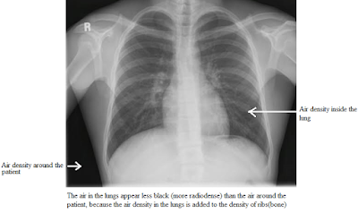
- Densities are additive
- Silhouette Sign
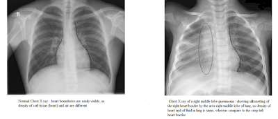
On an x-ray image, we can easily see the boundaries of tissue if its adjacent to the tissue with a different density like the boundary of heart shadow (grey) in a chest x-ray can easily be seen as its surrounded by air (black) which is radiolucent.
But on the other hand, if two structures with nearly the same density, then we can't see the boundaries of those structure as they got merged, in medical term it is known as silhouette sign.
Conclusion
- Radiology or in general said Medical imaging is an important part of clinical practice and even for the physiotherapist.
- This imaging technique plays an essential role to confirm the diagnosis and helps in the differential diagnosis.
- Knowledge and skills to interpret radiological images and reports are very important to have better communication with the other doctors/team members/co-workers and give the ability to take better decision to refer the patient if it’s not related to the field of physiotherapy.
- So, before directly jumping on the images to guessing which is this structure, we need to learn the basic concept related to imaging technique like how these images are formed? What do you mean by this black and grey colour in the x-ray? And many more questions are going to be answered in this blog series.
Resources
- Radiology Fundamentals: Introduction to Imaging and Technology fourth edition.
- Radiology Handbook: A Pocket Guide to Medical Imaging, J.S. Benseler, D.O.
- Lecture Notes Radiology, Third Edition, Pradip R. Patel.
- edX course: Applied anatomy of the locomotor system (Click here)
- Physio-pedia (Click here)



2 Comments
Easily understood
ReplyDeletewell explained
ReplyDeletePlease do not enter any spam link in the comment box