Introduction
We have studied the arthrokinematics of the knee joint in our post "how our knee joint work". But to move the knee joint, we need some active and passive stabilizers. In our previous post, we studied "passive knee stabilizers" but we also need "Active knee stabilizers" to move knee efficiently.
So, In this post, we are going to study "Active Knee Stabilizers" and how they guide Knee Arthrokinematics.
Active Knee Stabilizers
Active knee stabilizers are contractile structures, which stabilize our knee joint while movement like muscles, also known as dynamic stabilizers.
 |
Fig 1.1 Showing Front and back of the thigh |
Muscles of the knee joint
(A) Knee flexors
- There are 7 knee flexors as they cross the knee joint posteriorly are:
- Hamstrings(semimembranosus, semitendinosus, biceps femoris) sartorius, gracilis, popliteus and gastrocnemius. ( Fig 1.2 &1.3)
 |
| Fig 1.2 Front of thigh showing gracilus and sartorius |
 |
| Fig 1.3 Back of thigh showing Hamstring group |
- The semitendinosus, sartorius and gracilis form a common tendon, Pes anserinus(Goose foot). (Fig 1.4).
 |
| Fig 1.4 Saggital view of knee showing pes anserinus("Goose foot") |
- Rather than flexion, some of the muscles produce medial/lateral rotation or valgus/varus moments at the knee joint like:
- Semimembranosus, semitendinosus, sartorius, gracilis and popliteus also medially rotate the tibia on the femur.
- Biceps femoris, laterally rotate the tibia on the femur.
- Biceps femoris, lateral head of gastrocnemius and popliteus produce valgus moments at the knee (resist varus stress at the knee).
- Semimembranosus, semitendinosus, sartorius, gracilis and medial head of gastrocnemius produce varus moments at the knee (resist valgus stress at the knee).
(B) Knee extensors
- Quadriceps femoris (rectus femoris, vastus lateralis, vastus medialis, vastus intermedius).(Fig 1.5)
 |
Fig 1.5 Front of thigh showing Quadriceps femoris |
- The resultant pull of the vastus lateralis muscle is 35° laterally, whereas the resultant pull of the vastus medialis muscle is 40° medially, due to it's attachment to the medial and lateral aspects of patella respectively by the reticular fibres. (Fig 1.6)
 |
| Fig 1.6 Showing pull of Quadriceps femoris muscle include vastus medialis(VM), vastus lateralis(VL), vastus intermedius(VM), rectus femoris(RF (adapted from norkin 5th edition) |
- The pull of vastus intermedius muscle is parallel to the shaft of the femur.
- The net posterior compressive force due to vastus medialis (VM) and lateralis muscle(VL) is average 55°( Fig 1.7 )
 |
| Fig 1.7 Showing the posterior compressive force of VL and VM at 55° (adapted from norkin 5th edition) |
- The resultant pull of the quadriceps muscle is 10° to 15° in the lateral direction which is known as quadriceps angle(Q-angle). (Fig 1.8)
 |
| Fig 1.8 Front of thigh showing Q-angle |
- Patella:- The patella act as an anatomical pulley. It adds mechanical efficiency for quadriceps by increasing the moment arm of the quadriceps.
Clinical relevance:-
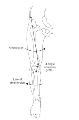
- Any alteration in alignment that increases the Q-angle is thought to increase the lateral patellar compressive forces and if force is more, may also lead to subluxate or dislocate the patella.
- Factors lead to increase Q- angle is:- Excessive genu valgum, Tight iliotibial band, Tight lateral retinaculum, medial femoral torsion, lateral tibial torsion, excessive or prolonged pronation in the foot. ( Fig 1.9)
 |
| Fig 1.9 Increased medial femoral (femoral anteversion) and tibial lateral torsion will result in a larger Q-angle and an increased lateral force on the patella |
- Q-angle is slightly greater in females than males, due to wider pelvis.
Role of muscles in arthrokinematic of Knee joint
- While weight-bearing knee flexion, semimembranosus and popliteus assist the menisci to deform posteriorly(Fig 1.10) so that menisci remain beneath the rounded femoral condyle as the condyles move on the relatively tibial plateau. The quadriceps contract eccentrically to counterbalance the knee flexion, and maintain the knee joint in equilibrium.
 |
Fig 1.10 Showing attachment of semimembranosus to the medial meniscus, which helps deform menisci posteriorly during knee flexion (adapted from norkin 5th edition) |
- While weight-bearing knee extension, the soleus and gluteus maximus assist the knee to produce extension (although they don't cross the knee joint).
How? By pulling the tibia posteriorly on femur due to contraction of the soleus muscle and by posterior shear of the femur on the tibia due to contraction of the gluteus maximus muscle. (Fig 1.11)
 |
Fig 1.11 Showing resultant pull of gluteus maximus and soleus assist in knee extension (adapted from norkin 5th edition) |
- While non-weight-bearing knee extension, the quadriceps contraction creates anterior shear of the tibia on the femur which is resisted by the active or passive forces like an anterior cruciate ligament.
- While non-weight-bearing knee flexion, hamstring contraction produce posterior shear of the tibia on femur and Popliteus assist the tibia to rotate medially to unlock the knee, posterior translation is resisted by Posterior cruciate ligament.
Summary
- Active knee stabilizers are contractile structure (dynamic stabilizers) like muscles.
- Knee flexors muscles like Hamstring, Sartorius, Gracilius, Popliteus.
- They also resist valgus and varus stress at the knee joint.
- Knee extensors muscles like Quadriceps femoris.
- Q-angle, the resultant pull of quadriceps femoris muscle.
- Increase in Q-angle, due to any malalignment of the body segment, lead to increase lateral compressive force on patella.
- Factors lead to increase Q-angle like medial femoral torsion, lateral tibial torsion, Excessive genu valgum, Excessive foot pronation.
- They also help in arthrokinamatic of the knee joint (weight-bearing/non-weight bearing).
Reference article
For more in details about the Q-angle, please click the reference link down below.
Resources
- Joint structure and function, a comprehensive analysis 5th edition by Pamela K. Levangie, Cynthia C. Norkin
- Kinesiology Of Musculoskeletal System, foundations for rehabilitation 2nd edition by Donald A. Neuman


3 Comments
Impressive .,🙏💐
ReplyDeletePlease sir /mamm, if it is possible you should have make 3D video on biomechanics to understand more eaisly for the students,
Because I still didn't see many videos regarding this .(please make if it possible , this my personal perseption)
Thank you
Thnk you and will surely bring 3-D video's, in later stage in our blog.
DeleteTill then keep in touch with us.
Ur effort seems here ...thnk u fo this
ReplyDeletePlease do not enter any spam link in the comment box